

The great explorer of the truth, the master-builder of human happiness no one rejects dislikes avoids pleasure itself because it is pleasure but because know who do not those how to pursue pleasures rationally encounter consequences that are extremely painful desires to obtain.
Read MoreVMA Technology
What Is Vertebral Motion Analysis?
A Modern Diagnostic Tool Built to Reveal What Static Imaging Misses
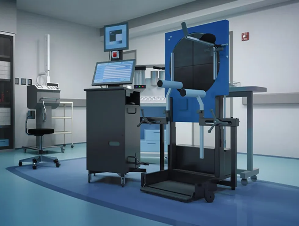
Vertebral Motion Analysis, or VMA®, is an FDA-cleared motion-based spine diagnostic that shows how the spine behaves in real time. Instead of taking still pictures like standard X-rays or MRIs, VMA captures continuous imaging as the patient gently bends forward, backward, and side to side.
Using a surgical-grade C-arm and proprietary software, VMA measures the movement of each vertebra with sub-millimeter accuracy. This reveals spinal ligament injuries and mechanical instability that are invisible on imaging at rest.
VMA replaces guesswork with objective, quantifiable data that supports better clinical decisions and more substantial legal proof.
Why Motion Matters
Most spinal injuries do not appear when the patient is lying flat or standing still. Pain, instability, and nerve irritation often occur only when the spine moves.
Traditional imaging is designed to capture anatomy, not function:
- X-rays show bones, not ligament integrity
- MRIs show soft tissue at rest, not segmental instability
- CT identifies fractures, not abnormal motion
- Flexion-extension X-rays show only two snapshots at end-range positions, while VMA solves the core gap in spinal diagnostics. It evaluates function, not just structure.
Guided Movement
The patient performs gentle, controlled bending while standing in the VMA. Movements are standardized and supported.
Real-Time Fluoroscopy
A surgical C-arm takes continuous images at eight frames per second throughout the bending cycle.
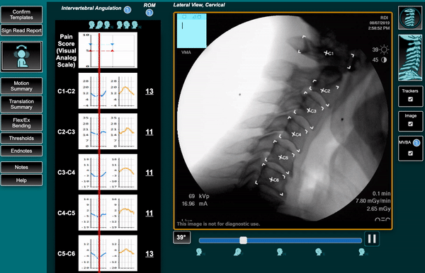
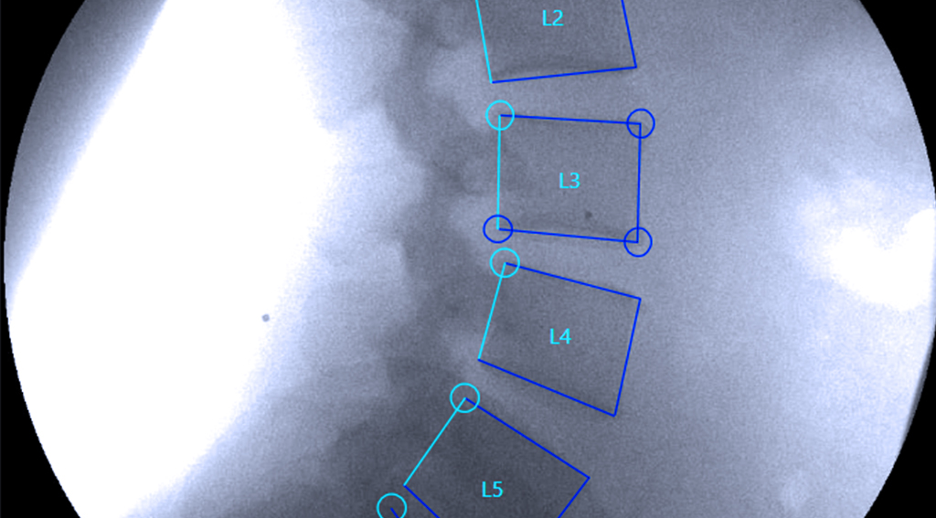
Quantitative Motion Analysis
Specialized software tracks and measures:
- Vertebral translation (sliding)
- Angular rotation
- Excessive motion patterns
- Segmental instability
- Motion asymmetry
- Abnormal load distribution
- Measurements are accurate within 0.5–0.7 mm, far beyond the sensitivity of standard imaging.
Independent Medical Interpretation
A board-certified radiologist analyzes the data and provides a formal diagnostic report with objective findings.
What VMA Reveals
VMA is uniquely capable of identifying:
- Spinal ligament injuries
- Alteration of Motion Segment Integrity (AOMSI)
- Segmental hypermobility or hypomobility
- Trauma-related instability
- Mechanical back or neck pain sources
- Whiplash-associated ligament injuries
- Subclinical or “hidden” injuries missed on MRI and X-ray
These injuries are among the most common causes of chronic pain after trauma, yet they are the most frequently overlooked.
The Technology Behind VMA
VMA is:
- FDA-cleared through 510(k) K172327
- Class II medical device (Product Code LLZ)
- Patented across nine motion-analysis innovations
- Validated with a 41% increase in sensitivity compared to traditional radiographs
- Backed by independent neuroradiology interpretations
- HIPAA-secure with cloud-based provider access
There is no sedation, no enclosed MRI tube, and no patient-controlled movement that might limit the accuracy of results.
Bring Clarity to the Spine
If your patient or client still hurts but their scans are “normal,” VMA can reveal the missing piece.
Refer a patient or request a case review today.